Sex-Related Cortical Alterations in Patients with Degenerative Cervical Myelopathy
Talia Oughourlian1,2,3, Chencai Wang2,3, Noriko Salamon3, Langston T. Holly4, Benjamin M. Ellingson2,3,4,5
1Neuroscience Interdisciplinary Graduate Program, David Geffen School of Medicine, University of California Los Angeles, Los Angeles, CA, USA; 2UCLA Brain Tumor Imaging Laboratory (BTIL), Center for Computer Vision and Imaging Biomarkers, David Geffen School of Medicine, University of California Los Angeles, Los Angeles, CA, USA; 3Dept. of Radiological Sciences, David Geffen School of Medicine, University of California Los Angeles, Los Angeles, CA, USA; 4Dept. of Neurosurgery, David Geffen School of Medicine, University of California Los Angeles, Los Angeles, CA, USA; 5Dept. of Psychiatry and Biobehavioral Sciences, David Geffen School of Medicine, University of California Los Angeles, Los Angeles, CA, USA
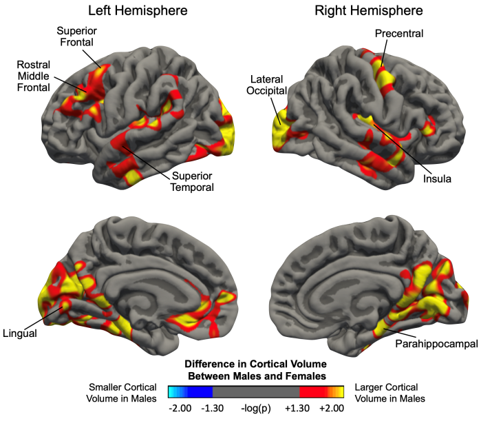
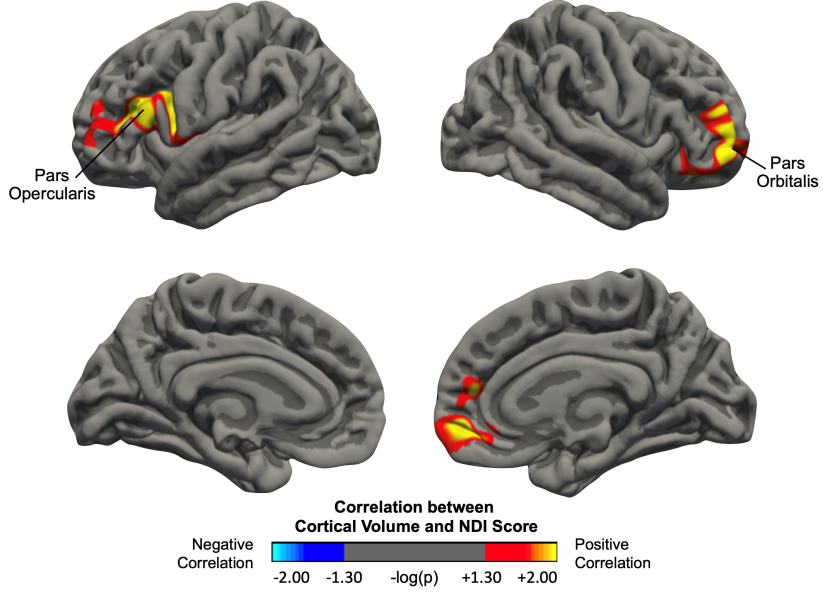
Introduction: Degenerative Cervical Myelopathy (DCM) is a progressive degenerative condition in which the cervical spinal cord becomes compressed leading to chronic neck pain or stiffness, weakness in the upper limbs, loss of fine motor skills, and/or limb dyscoordination. DCM is the most common spinal cord disorder in people over the age of 55. Studies have demonstrated that DCM patients undergo cerebral alterations including gray matter atrophy and white matter reorganization, yet sex-related cortical differences remain unknown. Sex-related morphological alterations have been observed in other chronic pain disorders including irritable bowel syndrome and migraine. We hypothesis patients with DCM will exhibit sex differences in cortical thickness and volume in sensorimotor and pain related regions. Methods: A total of 86 patients, 52 males and 34 females (mean age 59), with DCM were prospectively enrolled from 2016 to 2021 in a cross-sectional study involving brain imaging and evaluation of neck disability. Structural MRI images were acquired using a T1-weighted MP-RAGE sequence. The Neck Disability Index (NDI) questionnaire was used to measure the severity of neck pain and disability of patients (0 = no disability due to neck pain, 50 = complete disability). The NDI scores of the patient cohort ranged from 0 to 39 with an average of 12.6. FreeSurfer was used to perform cortical morphometric analyses, create general linear models, and perform statistical tests. All analyses controlled for subject age. The level of significance was set at p<0.05 with a false discovery rate (FDR) of 0.05. Results: Sex-related morphometric differences were observed within the DCM patient cohort. Specifically, whole brain analysis showed significantly thicker cortex in the left precuneus of male patients compared to female patients. Male patients also exhibited significantly larger cortical volume compared to female patients in regions including the left lingual gyrus, left superior frontal gyrus, left superior temporal gyrus, left rostral middle frontal gyrus, right lateral occipital cortex, right parahippocampal gyrus, right insular cortex, and right precentral gyrus (Fig. 1). Male patients exhibited a greater positive correlation between cortical volume and NDI score in the left pars opercularis and the right pars orbitalis compared to female patients (Fig. 2). Our results demonstrate that cortical volume varies as a function of NDI score in a sex-dependent way. Conclusion: In DCM patients, we observed sex-dependent cortical alterations in regions known to be involved in pain processing, including the precuneus, insula, superior frontal gyrus, rostral middle frontal gyrus, and precentral gyrus. Alterations in the precentral gyrus have been observed in other DCM studies and are suggested to be driven by somatosensory symptoms. Furthermore, males displayed a greater positive correlation between cortical volume and NDI in the pars opercularis and pars orbitalis suggesting that sex-dependent morphometric changes may be related to symptom severity. Changes in cortical thickness of the pars opercularis and pars orbitalis have been found in migraine patients compared to healthy controls. Our findings suggest differing cerebral alterations in males and females with DCM may influence disease symptomatology and may foster more effective treatment for patients suffering with DCM.
 |
 |
| Fig 1. A whole brain analysis comparing male and female DCM patients. Red-yellow color indicates larger cortical volume in male DCM patients, while blue-light blue indicates smaller cortical volume in male patients compared to female patients. Significant clusters were determined by thresholding based on statistical significance (p<0.05). | Fig 2. A whole brain analysis of DCM patients demonstrating a sex-dependent correlation between NDI score and changes in cortical volume. Red-yellow color indicates increased cortical volume with increased NDI score in males, while blue-light blue indicates decreased cortical volume with increased NDI score in males. Significant clusters were determined by thresholding based on statistical significance (p<0.05). |
Breakout Room: Oughourlian, Talia
View Poster: https://uclacns.org/symposium2021/16-Oughourlian-Talia.pdf
